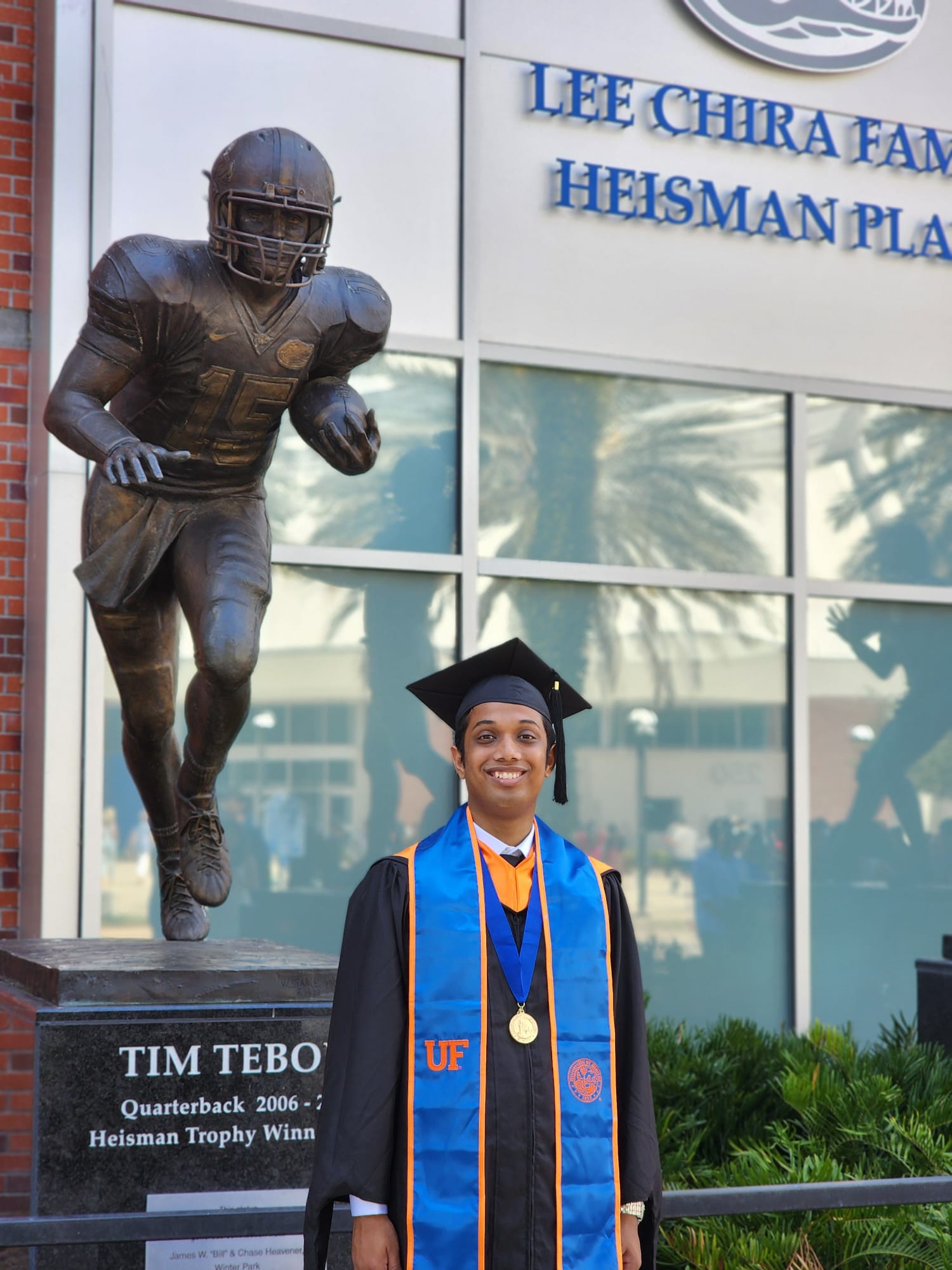
I graduated with a Masters degree in the ECE department of University of
Florida. Lost a lot of blood, broke several several bones and went through tremendous physical
and mental pain during this time. I am so proud that I did not quit.
July 24, 2023
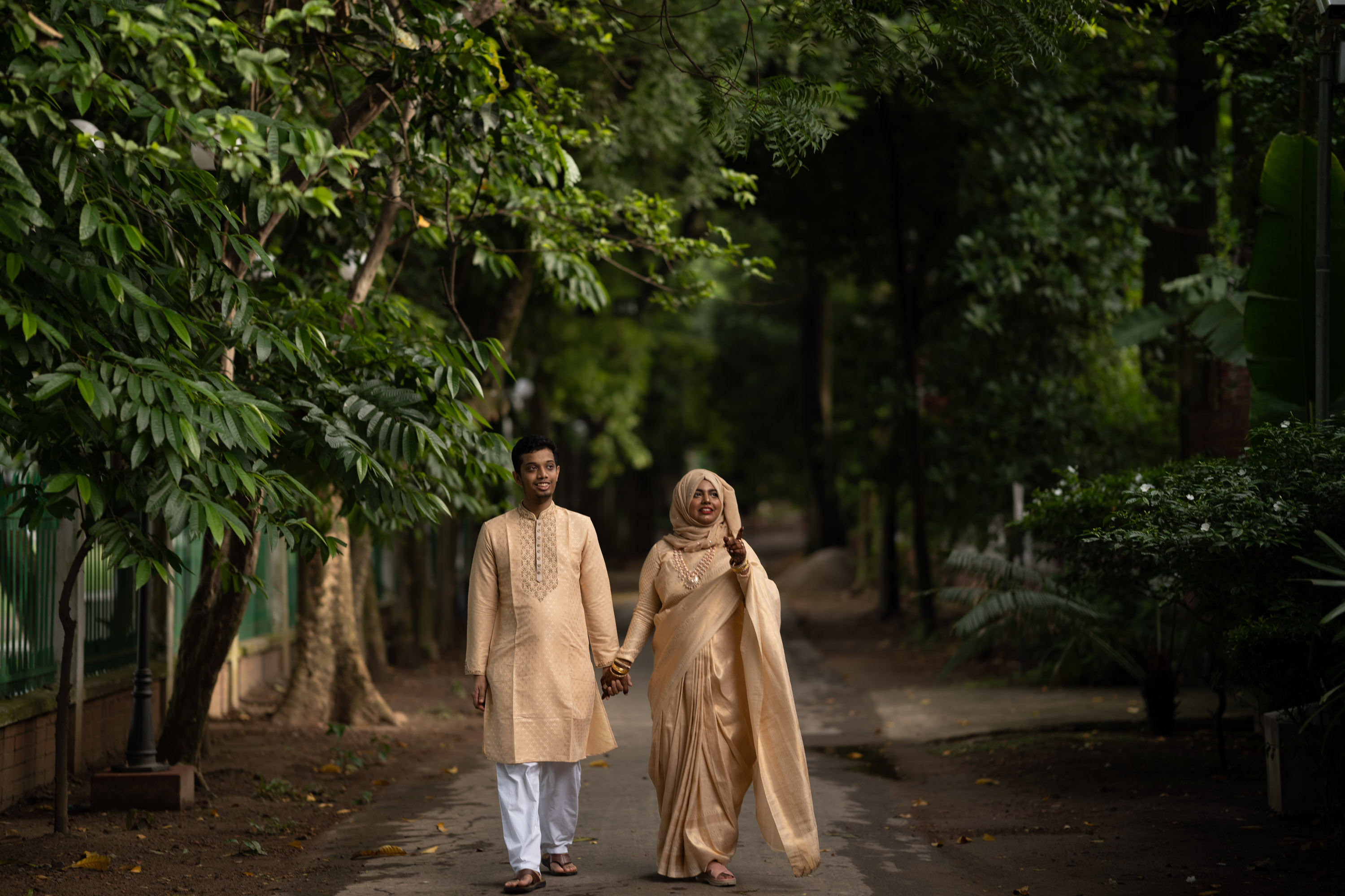
Married the Most Amazing Girl in the Whole World!
The title says it all! Her name is Trina, the love of my life.
January 2021
Joined FICS Lab as a Graduate Research Assistant
I started working under the supervision of Professor Navid Asadi in the FICS Lab in the department of ECE, University of Florida. I worked on projects
regarding physical assurance for AI and package security.
December 2019
Joined AI Samurai Japan Limited
AI Samurai Japan Limited is a sister concern of Chowagiken Company Limited Japan, a startup born
from Hokkaido University laboratories with the mission of "Putting research into practical use
and making it useful to society". They are focused on employing cutting-edge AI tools into
practical uses. I worked in this company as a Machine Learning Engineer.
February 2017
Graduated in EEE from BUET
I completed my graduation from the Electrical and Electronic Engineering
(EEE) department of Bangladesh University of Engineering and Technology (BUET) in February 2017.
August 2020
SIIM-ISIC Melanoma Classification 2020
The objective of this competition was to identify melanoma in images of
skin lesions. This was a binary classification problem where we had to predict whether the
melanoma condition in a provided image is benign or malignant. Our combined effort got us
253rd position (top 8%) on the Private Leaderboard among 3,314 teams. This was my
first bronze medal in any Kaggle competition and second medal overall. My detailed analysis and
codes are available here
in my GitHub repo. A lot of credit goes to my amazing team partners Mohammad Innat and Uday Kamal. Innat's well-explained solution is
available here.
February 2020
Web Application Project Got Accepted at SIIM
Intracranial Hemorrhage (ICH), which refers to bleeding inside the cranium,
is a serious health problem requiring rapid and intensive medical intervention. In case of an
emergency, it is necessary to urgently diagnose the subtype of IC from brain Computed Tomography
(CT) scans. However, in hospital emergency rooms in low-resource countries, a skilled
radiologist is almost non-existent, and an emergency medical officer has to make this urgent and
critical decision. This may lead to missed diagnosis, especially in the case of subarachnoid
hemorrhage which often shows subtle changes in the CT image. In this work, we have developed a
web-based AI tool for generating colorful heatmaps over the grayscale anatomical 2D CT image
slices identifying the possible regions of the subtype of acute IC. The tool can be used by
radiologists for AI-based assistance for making a more accurate and faster diagnosis. Our Web Application for
Intracranial Hemorrhage Detection got accepted at the Society for Imaging Informatics in Medicine (SIIM)
2020 Conference. Currently, the project is being supported by BrainStation 23. They have deployed a web tool
called RadAssist using our deep learning model. This
project was supervised by Dr. Taufiq
Hasan and Dr.
Paul Nagy.
October 2019
Joined mHealth Lab
mHealth Lab is working towards
developing biomedical signal processing and machine learning algorithms for remote health
monitoring, and prototyping innovative mHealth devices for improving people’s health, overall
well-being, and quality of life. This lab is founded and maintained by the Biomedical Engineering Department of BUET. I worked as a
Research Assistant (RA) there.
August 2019
APTOS 2019 Blindness Detection Competition
Diabetic retinopathy is a diabetes complication that affects eyes. It's
caused by damage to the blood vessels of the light-sensitive tissue at the back of the eye
(retina). It can be cured if treated at an early stage; otherwise, it could lead to permanent
blindness. Detection of the stage of diabetic retinopathy is a challenging task even for
doctors. Aravind Eye Hospital in India
collected thousands of retinopathy images and hosted this
competition for the detection of the level of DR in these images. In this competition, we
secured the position 37 on the Private and 55 among 2,943 teams on the Public leaderboard. Our
model scored 0.829 on the public LB and 0.926 on the Private LB. Codes are available here.
June 2019
Got Admitted to Masters Program
I got myself admitted to the Masters Program of EEE Department, BUET in
June 2019. I took courses on Modern Power System, Optical Fiber, Biomedical
Signal Processing, and Digital Image Processing.
December 2018
Published Research Article on Pathology Extraction from EHR
Extraction of relevant pathological terms from radiology reports is
important for correct image label generation and disease population studies. We published a
paper titled Pathology Extraction from Chest X-Ray Radiology Reports: A Performance Study
where we compared the performance of some known application program interfaces (APIs) for the
task of thoracic abnormality extraction from radiology reports. We explored several medical
domain-specific annotation tools like Medical Text Indexer (MTI) with Non-MEDLINE and MeSH On
Demand (MOD) options and generic Natural Language Understanding (NLU) API provided by the IBM
cloud. Our results showed that although MTI and MOD are intended for extracting medical terms,
their performance is worse compared to generic extraction APIs like IBM NLU. Finally, we trained
a DNN-based Named Entity Recognition (NER) model to extract the key concept words from radiology
reports. Our model outperformed the medical-specific and generic API performance by a large
margin. Our results demonstrated the inadequacy of generic APIs for pathology extraction tasks
and established the importance of domain-specific model training for improved results. The paper
is available here.
March 2017
Joined Semion Limited
Semion Limited is a startup founded by Dr. Khalid
Ashraf. The primary goal of this company is to create and promote services in the health
sector through digitization of patient and their associated data management. The digitization
process is backed up by tremendous technological advancement and customization through constant
research and development. They also develop artificial intelligence-based algorithms for
automating abnormality detection and assisting doctors. I worked as a Machine Learning
Researcher there.
February 2013 - June 2015
Worked as a Writer and Editor at Zero2Inf
I worked as a non-paid Writer and Editor at Zero2Infinity, a monthly science magazine, from March 2013 to
June 2015.